 (757) 622-2200
  (757) 622-2200
Right behind the pupil is the eye’s lens. It fine focuses the light directly onto the retina. The retina changes the light images into electrical impulses and sends them through the optic nerve to the processing center of the brain where vision becomes a vivid reality.
The Parts of an Eye
Eyes are responsible for about 75% of all that people perceive. The parts of an eye are designed to fulfill their specific roles in the amazing process of turning light into sight.
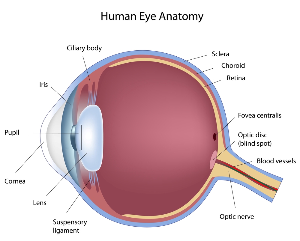
Cornea – The front “window” and main focusing element of the eye. It is made of a unique clear, but sturdy, tissue that allows light to pass through without distortion.
Sclera – The sturdy white tissue that forms the outer wall of most of the eye.
Choroid – A dark tissue under the white sclera that helps to trap light within the eye. Blood vessels within it serve to nourish much of the eye.
Iris – The visible colored part of the eye that expands and contracts to let the right amount of light in through its central opening for optimum vision.
Lens – The fine focusing element of the eye. Until a person is 40 to 50 years old, it can vary its curvature to focus at different distances.
Zonules – Flexible strands of tissue attached to the lens and the muscle of the ciliary body to facilitate the changes in lens curvature.
Ciliary Body – A rim of tissue inside the eye that consists of a muscle to change the curvature of the lens and a gland-like element that produces aqueous humor.
Aqueous Humor – A clear solution produced in the ciliary body to provide nutrients for the front tissues of the eye and to maintain internal eye pressure. It circulates through the front chambers of the eye and drains into the bloodstream.
Retina – A photosensitive membrane that lines the back inside wall of the eye. It captures the images of light, changes them into electrical impulses, and sends the messages to the processing center of the brain for interpretation into sight.
Macula – The central, indented part of the retina where light rays, ideally, come to a focal point. It is 100 times more sensitive that the other parts of the retina to allow clear vision of detailed objects.
Optic Nerve – The nerve that carries electrical impulses from the eye to the processing center of the brain.
Optic Disc – The place on the retina where the optic nerve joins the back of the eye. This area is not sensitive to light.
Vitreous Gel – A clear, gel-like substance that occupies the main cavity of the eye. It has no known function.


