Last Updated:
Our doctors are specially trained to handle all conditions of the retina and vitreous, including:
How the Eye Works: The Retina and the Macula
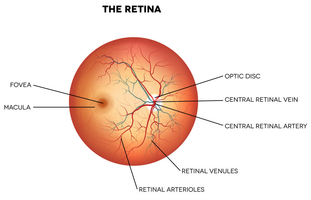
A thin layer of tissue called the retina covers the back inside wall of the eye. When the cornea and lens at the front of the eye focus light on the retina, a picture is taken and sent to the brain through the optic nerve to be interpreted. Thus, the retina is the “seeing tissue” of the eye.

The retina has two parts: the peripheral retina and the macula.
The “macula” is the center of the retina, while the large area surrounding the macula and comprising 95% of the retina is the “peripheral retina.” The peripheral retina allows us to see out of the corner of the eye, but is unable to see detail clearly. We cannot drive, read or see facial features with the peripheral retina.
Although small, the macula is one hundred times more sensitive to detail than the peripheral retina. The macula is required to see fine detail and it must be healthy to work properly.
Learn More Today
We invite you to learn more about our retina procedures, contact us now to schedule your consultation with one of our surgeons and learn about your options.
