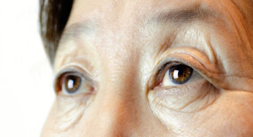
When an individual suffers a retinal detachment, a watery or wavy quality may be present. If the detachment happens in the peripheral retina, a shadow or “curtain” effect may be noticeable in the field of vision. When a retinal detachment occurs in the macula, reduced or distorted central vision can be a side effect.
Retinal problems that necessitate surgery are typically caused by the vitreous – a gel inside the eye that helps it maintain its spherical shape. The vitreous is attached to the retina, the macula, the optic nerve, and large retinal blood vessels.

Posterior Vitreous Detachment (PVD)
A Posterior Vitreous Detachment, or PVD, can occur when small groups of fluid develop inside the vitreous gel of the eye. These pockets of fluid can end up pulling on the retina, ultimately causing the vitreous to separate from the retina as well as the optic nerve. PVD is a very common occurrence and often results from the natural aging process.
Large spots in vision, or “floaters,” may be a symptom of PVD. These are typically caused by the vitreous becoming stringy and condensed. Flashes of light can also be symptomatic of PVD. These effects can occur as the vitreous pulls on the retina.
Retinal Tear and Vitreous Hemorrhage
If the vitreous pulls away from the retina in an area where the retina is weak, the retina may tear. If the retina tears across a retinal blood vessel, there will be bleeding into the vitreous. This is called a vitreous hemorrhage.
Retinal Detachment
When a retinal tear occurs, the liquid in the vitreous cavity may pass through the tear and get under the retina, collecting there and lifting the retina up off the back of the eye until it separates. Vision is lost wherever the retina becomes detached.

Treating Retinal Tears and Detachments
In Office Procedures
Laser treatment or cryotherapy, or both, may be used to seal a retinal tear and prevent a retinal detachment from happening. The laser is a beam of light that turns to heat when it hits the retina. The laser light is directed through a special lens. Cryotherapy is a means of freezing the part of the retina that needs to be treated, by placing a cryoprobe on the outside of the eye. Both laser treatment and cryotherapy seal the retina to the back wall of the eye.
Both laser surgery and cryotherapy are done on an outpatient basis. Patients may return to full activity in a short period of time.
Surgical Procedures
If the detachment is too large for laser treatment or cryotherapy alone, surgery is necessary to reattach the retina and avoid total loss of vision. There are three types of surgery for retinal detachment: scleral buckling and pneumatic cryopexy.
Scleral Buckling Surgery
Scleral Buckling Surgery is the traditional surgery for retinal detachment. The surgeon first treats the retinal tear with cryotherapy. A piece of silicone plastic or sponge is then sewn onto the outside wall of the eye (sclera) over the site of the retinal tear. This pushes the sclera in toward the retinal tear and holds the retina against the sclera until the tear is sealed.
Pneumatic Cryopexy
Pneumatic Cryopexy is another type of surgery that can be done for retinal detachments. Instead of placing a scleral buckle on the outside of the eye, the surgeon injects a gas bubble inside the vitreous cavity of the eye. The gas bubble then pushes the detached retina against the back wall of the eye to seal the retinal tear.
Vitrectomy
Vitrectomy is used when a retinal detachment is so complicated and severe that it cannot be treated with either standard scleral buckling surgery or pneumatic retinopexy. During the procedure, the vitreous is removed (thus the name “vitrectomy”) and replaced with clear fluid or with air that completely fills the eye. Over time, this fluid or air is absorbed and the eye replaces the vitreous with its own fluid.
Macular Holes
Macular holes are retinal holes in the center of the macula, caused by vitreous traction. The vitreous overlying the macula may contract and pull up the center of the macula, developing a tiny hole. With time, this hole may become larger. When a macular hole occurs, central vision is lost.

Once a macular hole develops, vitrectomy surgery may help. Our doctors will discuss the likelihood of it helping in your specific case.
Contact Virginia Eye Consultants
We invite you to learn more about our procedures for treating retinal detachment, retinal tears and macular holes. Contact us now to schedule your consultation with one of our surgeons and learn about your treatment options.
